
Hurthle Cell Thyroid Cancer: The Uncommon Diagnosis with Its Own Rules
Written by John P. Sabra, MD FACS
Updated September 2026
Educational only. This article is not medical advice. Always consult your physician about your individual situation.
Hurthle cell thyroid cancer, now also called oncocytic carcinoma, is an uncommon thyroid cancer that historically was grouped with follicular cancer but has been increasingly recognized as a distinct disease. It behaves somewhat more aggressively than typical follicular cancer, is less likely to absorb radioactive iodine, and warrants management by a team experienced specifically with this diagnosis. The basic principles are the same as other thyroid cancers, but several specific differences matter.
Hurthle cell cancer accounts for approximately 3 to 5% of thyroid cancers. The cells of origin are follicular cells that have undergone a distinctive transformation, becoming large with abundant pink cytoplasm packed with mitochondria. Under the microscope these cells have a characteristic appearance. Whether they form a benign Hurthle cell adenoma or a Hurthle cell carcinoma is determined the same way as for follicular cancer: by examining the capsule and blood vessels for invasion in the surgically removed specimen.
The Same Diagnostic Challenge as Follicular Cancer
Like follicular thyroid cancer, Hurthle cell cancer cannot be diagnosed on needle biopsy alone. The cells of a benign Hurthle cell adenoma look identical to those of a Hurthle cell carcinoma under the microscope. The distinction requires examining the capsule and blood vessels of the full specimen.
Biopsy of a Hurthle cell nodule typically returns a Bethesda IV result (follicular neoplasm, Hurthle cell type) and prompts the same diagnostic pathway as a follicular neoplasm. Molecular testing may be informative, and diagnostic lobectomy is often the definitive next step.
⚕ Clinical note: Molecular testing for Hurthle cell neoplasms has not been as informative as for typical follicular neoplasms. The genetic profile of Hurthle cell tumors is different, and some commercial molecular tests have lower specificity in this group. Discuss with your endocrinologist whether molecular testing changes the recommendation in your specific case before deciding to defer surgery based on a benign-classifier result.
How Hurthle Cell Cancer Differs from Typical Follicular Cancer
| Feature | Typical Follicular Cancer | Hurthle Cell Cancer |
|---|---|---|
| Lymph node spread | Uncommon (5 to 10%) | More common (10 to 25%) |
| Distant spread | Uncommon overall | More common, particularly to bone and lung |
| Iodine uptake | Generally good | Often poor; many tumors do not absorb iodine |
| Response to RAI therapy | Usually effective | Often less effective; alternative therapies considered earlier |
| Thyroglobulin as marker | Reliable | Usually reliable but somewhat less specific |
Surgical Treatment
Surgery is the foundation of treatment for Hurthle cell cancer. For minimally invasive Hurthle cell carcinoma confined to one lobe, lobectomy may be sufficient. For widely invasive disease, larger tumors, or any lymph node involvement, total thyroidectomy is standard. Lymph node dissection is performed when clinically indicated by imaging or operative findings.
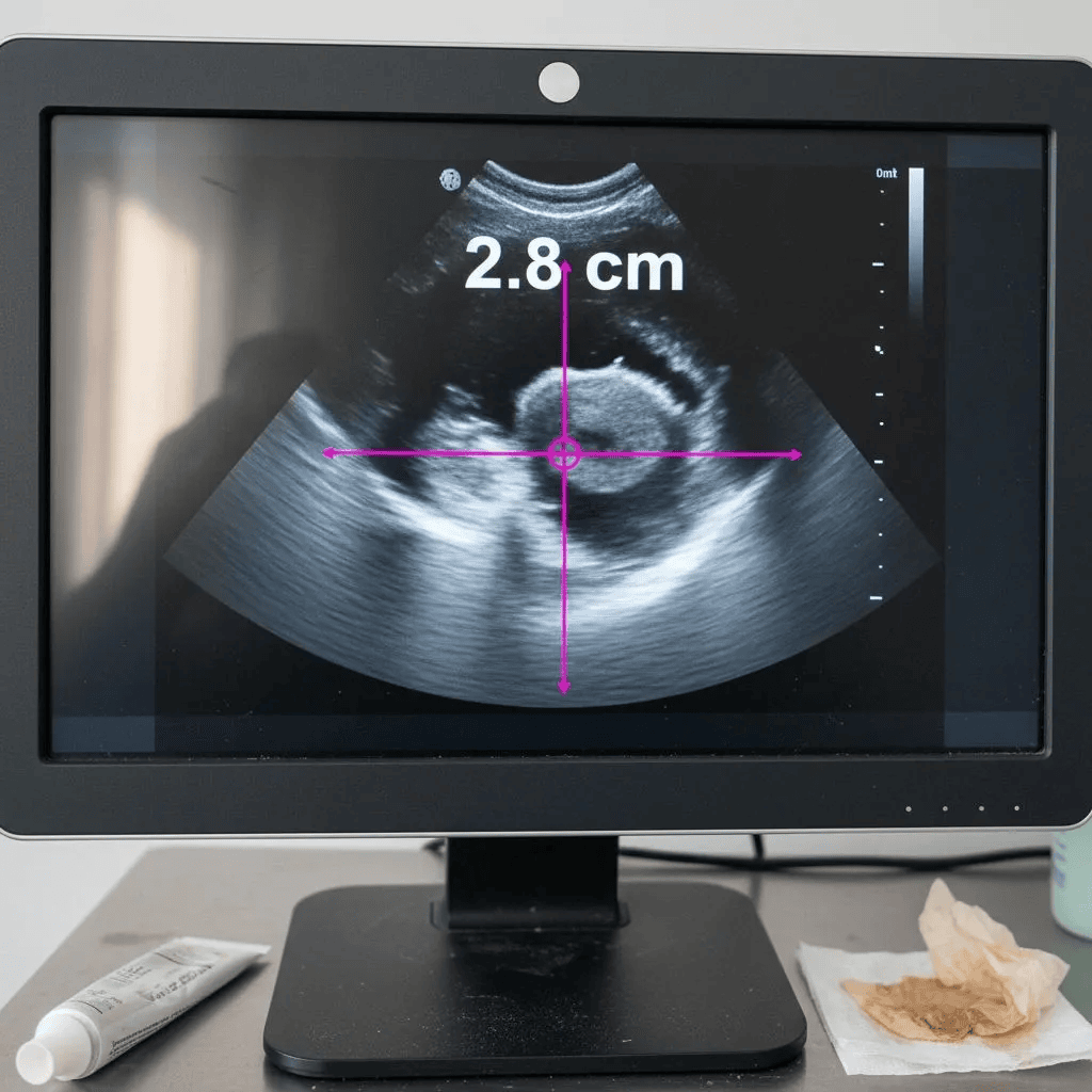
The somewhat higher rate of lymph node involvement in Hurthle cell cancer compared to typical follicular cancer is part of why preoperative neck ultrasound assessment is particularly important. A careful preoperative ultrasound by an experienced operator can identify suspicious lymph nodes that change the surgical plan from thyroidectomy alone to thyroidectomy plus central or lateral neck dissection.
The Role of Radioactive Iodine
Radioactive iodine therapy is less consistently effective in Hurthle cell cancer than in typical follicular cancer because Hurthle cells often do not absorb iodine as readily. This does not mean RAI is never useful. It is still considered in higher-risk patients, particularly when there is concern for residual or metastatic disease. The dose may be higher, and the response is less predictable.
For Hurthle cell cancer that does not respond to RAI, targeted therapy with tyrosine kinase inhibitors has emerged as an important option. Drugs such as lenvatinib and sorafenib have been used in advanced iodine-refractory disease and can produce clinical responses.
Surveillance
Surveillance after Hurthle cell cancer treatment follows the same general framework as other differentiated thyroid cancers, using thyroglobulin and neck ultrasound. Because distant recurrence is somewhat more common, imaging beyond the neck (chest CT, bone imaging, FDG-PET) may be considered in selected higher-risk patients. The surveillance schedule is individualized.
The Bottom Line
Hurthle cell thyroid cancer is uncommon, distinct from typical follicular cancer in several important ways, and best managed by a team experienced with the diagnosis. Surgery is the foundation of treatment. Radioactive iodine is less consistently effective. Targeted therapy is an established option for iodine-refractory disease. Outcomes for minimally invasive Hurthle cell cancer are generally favorable; widely invasive or metastatic disease requires more intensive management.
If your pathology returns as Hurthle cell carcinoma, seek evaluation at a center experienced with this specific diagnosis, and ask about the team’s approach to iodine therapy and targeted therapy.
This article is intended for patient education only. It does not constitute medical advice and does not replace a consultation with your physician. Individual patient circumstances and clinical judgment determine the appropriate evaluation and management for every patient.
More from the Thyroid Cancer series



Ready to Talk to a Specialist?
A thyroid specialist can review your specific results, answer your questions, and help you understand exactly what your next steps should be. The ATLAS network connects you with experienced thyroid and parathyroid expert physicians who focus on thyroid disease.
Find a Thyroid Specialist Near You →