Follicular Thyroid Cancer: Why Needle Biopsy Cannot Diagnose It and What Comes Next
Written by John P. Sabra, MD FACS
Updated September 2026
Educational only. This article is not medical advice. Always consult your physician about your individual situation.
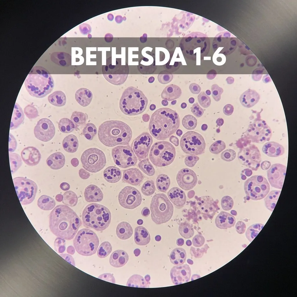
Follicular thyroid cancer is the second most common form of thyroid cancer, and it has a feature that makes it unique among thyroid cancers: it cannot be diagnosed on a fine needle biopsy. The cells of a benign follicular adenoma look identical to the cells of a follicular carcinoma under the microscope. The only way to distinguish the two is by examining the entire tumor after it has been removed. This is one of the most counterintuitive facts about thyroid cancer, and one of the most important to understand if your biopsy came back as Bethesda IV.
Follicular thyroid cancer accounts for approximately 10 to 15% of thyroid cancer diagnoses in the United States. Like papillary thyroid cancer, it arises from the follicular cells of the thyroid and is considered a differentiated thyroid cancer. It tends to behave somewhat differently from papillary cancer in two important ways: it spreads less often to lymph nodes, and it spreads more often through the bloodstream to lungs or bones. Even with those differences, survival rates remain favorable, and most patients diagnosed with follicular thyroid cancer do well long-term.
The diagnostic challenge of follicular cancer is the central reason it deserves special attention. If your biopsy result was Bethesda IV (follicular neoplasm), you are facing a particular kind of uncertainty: a nodule that may or may not be cancer, with the only definitive answer coming from a surgical pathologist after part of your thyroid has been removed. Understanding why this is the case, and what the options are, makes the next decision easier.
What Makes Follicular Cancer Different
The hallmark of follicular thyroid cancer is capsular invasion or vascular invasion by the cancer cells. A follicular adenoma is a benign tumor encased in a thin capsule; the cells stay within that capsule. A follicular carcinoma looks the same under the microscope but has broken through the capsule into the surrounding thyroid tissue or invaded small blood vessels. That difference is what makes one a benign growth and the other a cancer.
A needle biopsy samples only a small portion of cells from the center of the nodule. It cannot examine the capsule itself, or assess if cells have invaded the capsule. It cannot examine the blood vessels. So when the pathologist looks at a follicular biopsy specimen and sees follicular cells, the report comes back as Bethesda IV: follicular neoplasm. The malignancy risk associated with this category is 25 to 40%, meaning the majority of these nodules turn out to be benign, but a meaningful minority will be cancer.
The Bethesda IV result does not mean you have cancer. It means the cells could not distinguish between a benign adenoma and a cancer, and a more definitive answer requires examining the full nodule, or assessing further with genetic testing. Roughly 60 to 75% of Bethesda IV nodules turn out to be benign when removed.
Molecular Testing as the Bridge Between Biopsy and Surgery
Over the past decade, molecular testing has become an extremely important tool for managing Bethesda III and IV nodules. Tests such as Afirma and ThyroSeq analyze the genetic profile of the biopsied cells and can often reclassify a follicular neoplasm as likely benign or likely malignant. This genetic testing cannot give an answer with 100% certainty, but it comes close.
For a patient with a Bethesda IV result and a benign molecular profile, the malignancy risk falls below 5%, and surveillance becomes a very reasonable alternative to surgery. For a patient with a suspicious molecular profile, the result strengthens the case for proceeding with surgery and may even guide the extent of the operation. Molecular testing does not replace surgery in every case, but it has substantially reduced the number of diagnostic operations performed on what turn out to be benign nodules.
When Surgery Is the Right Next Step
For Bethesda IV nodules that are either not tested with molecular testing or that test suspicious, the standard next step is a diagnostic thyroid lobectomy. The lobectomy removes the half of the thyroid containing the nodule. The full specimen is then examined by a pathologist, who can assess the capsule and blood vessels and determine whether the nodule is a benign adenoma or a follicular carcinoma.
| Final Pathology Result | What It Means | What Comes Next |
|---|---|---|
| Follicular adenoma | Benign growth. No cancer present. | No further surgery needed. Surveillance of the remaining thyroid lobe. |
| Minimally invasive follicular carcinoma | Cancer with only capsular invasion, no significant vascular invasion. | Lobectomy is often sufficient definitive treatment. Surveillance ultrasound and thyroid labs. |
| Widely invasive follicular carcinoma | Cancer with extensive capsular or vascular invasion. | Completion thyroidectomy and possible radioactive iodine therapy. |
How Follicular Cancer Behaves When It Is Present
Follicular thyroid cancer tends to spread differently from papillary cancer. Lymph node involvement is uncommon, occurring in roughly 5 to 10% of cases. Distant spread through the bloodstream to lungs or bones is more common with follicular cancer than with papillary cancer, but is still relatively uncommon overall.
The distinction between minimally invasive and widely invasive follicular carcinoma is one of the most important findings in the pathology report. Minimally invasive disease has a 10-year survival rate of greater than 95% and is usually well-treated with lobectomy alone in patients without other risk factors. Widely invasive disease behaves more aggressively, has a meaningfully higher recurrence rate, and almost always warrants total thyroidectomy plus radioactive iodine.
The pathology of follicular carcinoma matters more than the size of the tumor in many cases. A 4 cm minimally invasive follicular cancer may need less aggressive treatment than a 2 cm widely invasive one. Make sure your surgeon and endocrinologist have reviewed the full pathology report, including the assessment of capsular and vascular invasion, before any decisions about additional surgery or radioactive iodine therapy.
The Special Case of Hurthle Cell Carcinoma
Hurthle cell carcinoma (also called oncocytic carcinoma) is a subtype of cancer historically grouped with follicular cancer. Hurthle cells are large follicular cells with abundant pink cytoplasm packed with mitochondria, giving them a distinctive appearance. The diagnostic problem is identical to that for follicular cancer: a needle biopsy cannot reliably distinguish a benign Hurthle cell adenoma from a Hurthle cell carcinoma, and a diagnostic lobectomy is often required.
Hurthle cell carcinomas tend to behave somewhat more aggressively than typical follicular carcinomas, are less likely to absorb radioactive iodine, and warrant management by surgeons and endocrinologists experienced with the diagnosis. If your pathology returns as Hurthle cell carcinoma, ask whether your team treats this diagnosis regularly.
Surveillance After Treatment
Surveillance after follicular thyroid cancer treatment is similar to surveillance after papillary cancer treatment. Patients who have had total thyroidectomy are followed with thyroglobulin levels, neck ultrasound, and TSH lab monitoring on a schedule that becomes less frequent over time. Patients treated with lobectomy alone for low-risk minimally invasive disease are followed with periodic ultrasound of the remaining thyroid lobe and thyroid function tests.
Because follicular cancer is more likely than papillary cancer to spread to lungs or bones, surveillance imaging with scans beyond the neck may be considered in higher-risk patients. The specifics depend on the final pathology and the judgment of the treating team.
The Bottom Line
Follicular thyroid cancer is the second most common type of thyroid cancer and is unique in that it cannot be diagnosed with a fine needle biopsy alone. A Bethesda IV biopsy result reflects this diagnostic limitation, not a cancer diagnosis. Genetic testing is an excellent option that can give a “likely benign” diagnosis with 95% confidence, and spare many patients from surgery. If surgery is deemed necessary in some patients, most Bethesda IV nodules turn out to be benign once removed and examined in full. For those that are confirmed as follicular carcinoma, the outlook for minimally invasive disease is excellent, and treatment is increasingly being tailored to the pathology rather than a one-size-fits-all approach.
If your biopsy came back as Bethesda IV, ask whether molecular testing has been performed and what the result is, and ask your surgeon whether a diagnostic lobectomy is being recommended on the basis of those results.
This article is intended for patient education only. It does not constitute medical advice and does not replace a consultation with your physician. Individual patient circumstances and clinical judgment determine the appropriate evaluation and management for every patient.
More from the Thyroid Cancer series



Ready to Talk to a Specialist?
A thyroid specialist can review your specific results, answer your questions, and help you understand exactly what your next steps should be. The ATLAS network connects you with experienced thyroid and parathyroid expert physicians who focus on thyroid disease.
Find a Thyroid Specialist Near You →