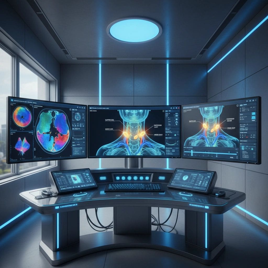
Incidental Thyroid Nodule: Found on a CT or MRI You Had for Something Else
Written by John P. Sabra, MD FACS
Updated September 2026
Educational only. This article is not medical advice. Always consult your physician about your individual situation.
A thyroid nodule found by accident on a scan done for another reason is called an incidental thyroid nodule, or an “incidentaloma”. It is one of the most common findings in modern radiology, showing up on chest CT’s, carotid ultrasounds, neck MRI’s, and PET scans that are done for completely unrelated reasons. Most are benign. Few require any treatment. But the appropriate next steps depend on size and on the type of scan that found it.
Incidental thyroid nodules are common because modern imaging is sensitive enough to detect anatomy that older generations of scans missed entirely. Approximately 20% of contrast-enhanced chest or neck CT’s include an incidentally found thyroid nodule. The phenomenon is similar across MRI, PET, and ultrasound of nearby structures. The overall malignancy risk of an incidental nodule is approximately the same as any thyroid nodule, around 5 to 10%, depending on the specific features.
The Right First Step Is Almost Always a Dedicated Thyroid Ultrasound
The CT, MRI, or PET that found the nodule was not designed to characterize the thyroid. The resolution is lower than thyroid ultrasound, the features cannot be assessed with the same precision, and a nodule classification cannot be assigned. A dedicated thyroid ultrasound is the right next test for the great majority of incidental nodules. It defines the size, the precise location, the internal features, and the appearance of lymph nodes.
⚕ Clinical note: Do not rely on the description of the nodule from the original CT or MRI report alone. The original radiologist may note size and basic features, but the level of detail needed to make decisions about biopsy or surveillance comes from a focused thyroid ultrasound with nodule risk classification.
Size Thresholds for Pursuing Evaluation
Not every incidentally found nodule requires dedicated ultrasound. Several professional societies have developed guidelines on which incidental nodules warrant further imaging.
| Patient Profile | Dedicated Ultrasound Recommended at |
|---|---|
| Adult under 35 years old | Any incidentally noted nodule. |
| Adult 35 years or older | 1.0 cm or larger. |
| Any age with concerning features on cross-sectional imaging | Any size. |
| PET-positive thyroid finding | Any size. PET-positive thyroid foci have a higher malignancy risk and warrant prompt evaluation. |
The Special Case of PET-Positive Thyroid Findings
An incidental thyroid finding on a PET scan is different from an incidental finding on CT or MRI. PET highlights areas of increased metabolic activity. A focal area of increased uptake within the thyroid carries an estimated malignancy risk of 25 to 35%, substantially higher than the general thyroid nodule population. Diffuse increased uptake throughout the thyroid usually represents thyroiditis (such as Hashimoto disease) rather than cancer, but focal uptake should always be evaluated with dedicated thyroid ultrasound and almost always with biopsy if a discrete nodule is found.
When No Further Workup Is Needed
For very small incidental nodules in older adults (typically under 1 cm in patients over 35), most professional societies do not recommend further workup. The reasoning is that even if a small papillary cancer were present, the great majority would never progress to clinical significance, and the morbidity of additional testing and procedures outweighs the small potential benefit.
-
Adult over 35, incidental nodule under 1 cm, no concerning features: No specific follow-up, but discuss getting a follow up ultrasound with your physician.
-
Adult over 35, incidental nodule 1 to 1.5 cm, no concerning features: Selective evaluation based on patient factors, typically an ultrasound follow-up
-
Any patient with small simple cystic nodules: No further workup needed.
⚕ Clinical note Patients with a history of head or neck radiation, a family history of thyroid cancer, or features of MEN syndromes are evaluated more thoroughly even for small incidental findings. The general size thresholds above do not apply in these higher-risk populations.
What to Bring to Your Next Visit
-
The original scan report: The radiology report from the CT, MRI, or PET that found the nodule.
-
The images if possible: A disc or electronic copy of the images can be useful for the doctor performing the dedicated thyroid ultrasound to compare.
-
Your medical history: Family history of thyroid cancer, any past head or neck radiation, prior thyroid imaging if available.
The Bottom Line
An incidentally found thyroid nodule is common, almost always benign, and rarely an emergency. The right first step for most incidental nodules of sufficient size is a dedicated thyroid ultrasound with nodule risk classification, followed by appropriate biopsy or surveillance based on the result. Very small incidental nodules in older adults often need no specific workup. PET-positive incidental thyroid findings are a specific exception that always warrants evaluation.
Schedule a dedicated thyroid ultrasound with the report and images from your original scan, and use the resulting classification to guide what comes next in a discussion with your doctor.
This article is intended for patient education only. It does not constitute medical advice and does not replace a consultation with your physician. Individual patient circumstances and clinical judgment determine the appropriate evaluation and management for every patient.
More from the Ultrasound series




Ready to Talk to a Specialist?
A thyroid specialist can review your specific results, answer your questions, and help you understand exactly what your next steps should be. The ATLAS network connects you with experienced thyroid and parathyroid expert physicians who focus on thyroid disease.
Find a Thyroid Specialist Near You →