CT, MRI, and PET Imaging for Thyroid Disease: When Do You Actually Need Them?
Written by John P. Sabra, MD FACS
Updated September 2026
Educational only. This article is not medical advice. Always consult your physician about your individual situation.
Ultrasound is the foundation of thyroid imaging. It does the great majority of the diagnostic work, including assessing nodules, lymph nodes, and gland size. CT, MRI, and PET each have specific roles that ultrasound cannot fill. Knowing when each is the right test and what it adds is part of how the modern workup is organized.
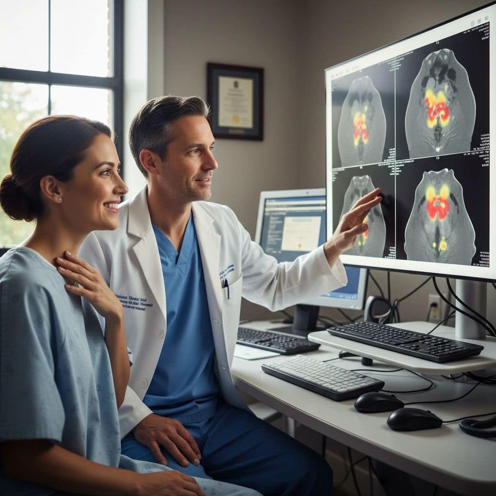
Each imaging modality answers a different question. Ultrasound is best at characterizing the thyroid gland itself and the lymph nodes immediately around it. CT and MRI provide three-dimensional anatomy useful for surgical planning and for evaluating disease that extends beyond the thyroid. PET shows metabolic activity and is most useful in selected thyroid cancer surveillance situations.
When CT Is the Right Test
-
Substernal extension of thyroid disease: To determine how far into the chest the thyroid extends, which affects the surgical approach.
-
Large goiter compressing the airway: To evaluate the airway and surrounding structures.
-
Advanced thyroid cancer with suspected invasion: To assess invasion of the trachea, esophagus, or vascular structures.
-
Mediastinal lymph nodes: For thyroid cancer suspected to involve lymph nodes in the chest.
-
Pre-operative planning for complex cases: Reoperation, locally advanced disease, or unusual anatomy.
⚕ Clinical note: For thyroid cancer patients about to undergo radioactive iodine therapy, CT scans with iodinated contrast should be timed carefully. The iodine in CT contrast saturates the thyroid and reduces the effectiveness of subsequent RAI. Most protocols delay RAI for 4 to 8 weeks after iodinated contrast, or use non-contrast CT or MRI instead when imaging is needed close to planned RAI.
When MRI Is the Right Test
-
Pregnancy: When advanced imaging is needed and radiation should be avoided.
-
Contrast allergy or kidney disease: When iodinated CT contrast is contraindicated.
-
Soft tissue invasion assessment: MRI shows soft tissue planes better than CT in some cases.
-
Brain or spine surveillance in advanced thyroid cancer: When metastatic disease is being evaluated.
-
Selected parathyroid localization: When CT and ultrasound have been non-localizing.
When PET Is the Right Test
FDG-PET (fluorodeoxyglucose PET) is not part of the routine workup for thyroid nodules or initial thyroid cancer staging. Its role is in specific situations during thyroid cancer surveillance, particularly for differentiated thyroid cancer that has become iodine-refractory.
-
Rising thyroglobulin with a negative iodine scan and other imaging: PET can identify disease not seen elsewhere, particularly when thyroglobulin is above 10 ng/mL.
-
Iodine-refractory differentiated thyroid cancer: When the cancer no longer absorbs iodine, PET may identify residual disease.
-
Aggressive thyroid cancers: Anaplastic, poorly differentiated, and some Hurthle cell cancers are PET-avid.
-
Evaluation of suspicious findings on other imaging: Selected use to clarify whether a finding is metabolically active.
⚕ Clinical note: Low-risk differentiated thyroid cancer does not typically take up FDG well, so a negative PET scan in a low-risk patient does not rule out disease. On the flip side, an incidental focal uptake in the thyroid on a PET scan done for another reason has a 25 to 35% chance of being thyroid cancer and warrants evaluation with dedicated thyroid ultrasound.
Side-by-Side Summary
| Modality | Best For | Trade-Off |
|---|---|---|
| Ultrasound | Thyroid nodules, lymph nodes, gland anatomy. | Cannot see into chest or deep structures. |
| CT | Substernal extension, mediastinal disease, advanced cancer. | Radiation, iodine contrast may delay RAI. |
| MRI | Pregnancy, contrast contraindication, soft tissue detail. | Limited availability, longer exam time, claustrophobia issues. |
| PET | Iodine-refractory cancer, rising Tg with negative other imaging. | Not useful for routine thyroid cancer or low-risk disease. |
The Bottom Line
Ultrasound is the foundation of thyroid imaging and does the majority of the diagnostic work. CT is the standard for substernal disease, advanced cancer staging, and complex surgical planning. MRI is the alternative when CT is contraindicated. PET has specific use in iodine-refractory differentiated thyroid cancer and in rising thyroglobulin with negative other imaging. Each test answers a different question, and ordering the right test means matching the question to the modality.
If your physician orders advanced imaging beyond ultrasound, ask what specific question the test is meant to answer and how the result will change the treatment plan.
This article is intended for patient education only. It does not constitute medical advice and does not replace a consultation with your physician. Individual patient circumstances and clinical judgment determine the appropriate evaluation and management for every patient.
More from the Technology and Tools series

Ready to Talk to a Specialist?
A thyroid specialist can review your specific results, answer your questions, and help you understand exactly what your next steps should be. The ATLAS network connects you with experienced thyroid and parathyroid expert physicians who focus on thyroid disease.
Find a Thyroid Specialist Near You →