4D-CT for Parathyroid Disease: When Standard Imaging Is Not Enough
Written by John P. Sabra, MD FACS
Updated September 2026
Educational only. This article is not medical advice. Always consult your physician about your individual situation.
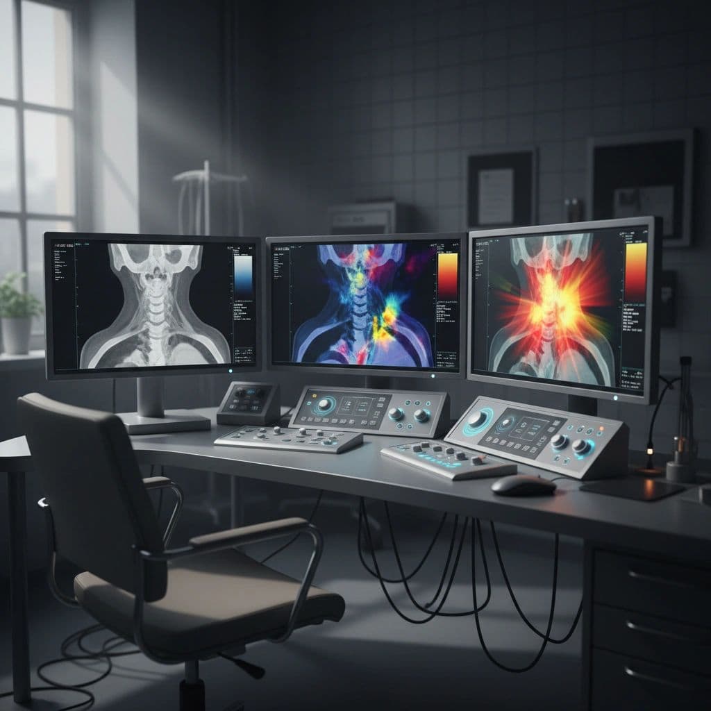
4D-CT is a specialized CT scan technique developed specifically for locating abnormal parathyroid glands. When ultrasound and sestamibi scan have not produced a clear answer, 4D-CT often does. It has become an essential tool for the more difficult parathyroid cases and for patients facing reoperation. Knowing when it is appropriate and what it can and cannot show completes the imaging picture.
The “4D” in 4D-CT refers to four dimensions: the three spatial dimensions of a standard CT scan plus a fourth dimension of time. Images are obtained at three or four different time points after contrast injection. Abnormal parathyroid glands show a characteristic pattern of contrast uptake and washout that differs from normal lymph nodes, thyroid tissue, or other structures. That temporal pattern is what makes 4D-CT more accurate than a single CT scan.
The sensitivity of 4D-CT for parathyroid adenoma is approximately 85 to 95% in published series at experienced centers. This is meaningfully higher than the 70 to 80% sensitivity of sestamibi scan. The trade-off is higher radiation exposure and the need for iodinated contrast.
When 4D-CT Is the Right Test
-
Negative sestamibi scan and negative ultrasound with biochemical primary hyperparathyroidism: 4D-CT often finds the adenoma when the initial imaging did not.
-
Reoperative parathyroid surgery: After a failed first operation, 4D-CT is essential for localizing the remaining abnormal tissue. Surgical landmarks are disrupted after the initial surgery, which makes other imaging less reliable.
-
Suspected ectopic parathyroid: 4D-CT covers the neck and upper chest and can identify glands in unusual locations.
-
Multi-gland disease evaluation: 4D-CT can sometimes identify multiple abnormal glands when other imaging shows only one.
⚕ Clinical note: 4D-CT is not typically the first imaging test for primary hyperparathyroidism, because of its higher radiation dose. The standard initial imaging is ultrasound combined with sestamibi scan. 4D-CT is reserved for when those tests have not adequately localized the disease, or for reoperative cases where its higher resolution and broader anatomic coverage are essential.
What 4D-CT Shows
-
Three-dimensional anatomy of the neck and upper chest: A complete map of where the abnormal gland sits relative to the thyroid, esophagus, trachea, and vascular structures.
-
Pattern of contrast enhancement over time: Distinguishes parathyroid from lymph node or thyroid tissue.
-
Identification of ectopic glands: Glands in the chest, behind the esophagus, within the thyroid, or in other unusual locations.
-
Surgical anatomy: The surgeon can plan the approach in detail before entering the operating room.
Limitations
-
Radiation exposure: Higher than sestamibi scan. Not a routine first-line test in younger patients.
-
Iodinated contrast: Some patients have a contrast allergy or kidney problems that limit its use.
-
Interpretive expertise required: 4D-CT reading requires a radiologist familiar with parathyroid imaging. Centers without this expertise may produce less accurate reads.
-
Cost: More expensive than sestamibi scan. Insurance coverage varies.
-
Small or multi-gland disease can still be missed: No imaging is perfect.
When the Imaging Approach Goes Beyond 4D-CT
A small subset of patients have biochemical parathyroid disease that all standard imaging fails to localize. For these patients, additional options include MRI (radiation-free, useful in pregnancy and patients with contrast contraindications), PET imaging with specific tracers (available at some research centers), and selective venous sampling (an invasive test that measures PTH levels from veins in different regions of the neck to identify the side or area of disease).
Even with negative imaging across all modalities, surgery for biochemical primary hyperparathyroidism remains appropriate. Experienced parathyroid surgeons can find the abnormal gland during the operation with high success rates using systematic exploration techniques. The role of imaging is to make surgery easier, not to make it possible.
⚕ Clinical note: 4D-CT reading requires specific experience. The same scan read at a center with a high-volume parathyroid program may yield a clear localization, while the same scan read at a general radiology practice may be reported as negative. If your 4D-CT is read as negative and your surgeon recommends reoperation or extensive exploration, getting the images formally reviewed at a center with parathyroid expertise is sometimes informative.
The Bottom Line
4D-CT is a specialized imaging test with high sensitivity for parathyroid localization. It is the next step when ultrasound and sestamibi scan have not produced a clear answer, and it is essential for reoperative parathyroid surgery. The trade-offs are higher radiation, contrast use, and the need for experienced interpretation. Even with all imaging negative, surgery for biochemical disease is still appropriate in experienced hands.
If your initial parathyroid imaging is non-localizing, ask whether 4D-CT is available locally and whether the reading radiologist has experience with parathyroid imaging.
This article is intended for patient education only. It does not constitute medical advice and does not replace a consultation with your physician. Individual patient circumstances and clinical judgment determine the appropriate evaluation and management for every patient.
More from the Parathyroid Disease series



Ready to Talk to a Specialist?
A thyroid specialist can review your specific results, answer your questions, and help you understand exactly what your next steps should be. The ATLAS network connects you with experienced thyroid and parathyroid expert physicians who focus on thyroid disease.
Find a Thyroid Specialist Near You →